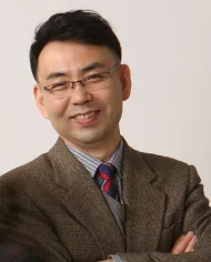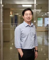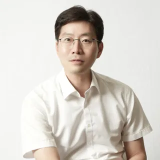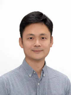
조형준 교수 사진

UNIST바이오메디컬공학과

MRI 뉴로영상 연구실

조형준 교수 (Hyung Joon Cho)

연구실 소개

이 연구실은 동적 조영증강 MRI와 확산 MRI, PET를 활용해 종양의 관류·저산소 상태와 종양 미세환경을 영상화하고, 영상 지표와 종양 조직을 비교하는 연구를 합니다. 또한 SPION을 이용해 MRI에서 T1·T2* 이중 대비 영상을 획득하고, 이를 통해 혈관과 조직을 관찰하는 방법을 연구합니다.

AI가 연구실 홈페이지를 바탕으로 생성한 연구실 소개예요

연구실 홈페이지의 소개

Imaging tumor microenvironment (viable, necrotic and hypoxic region) can provide prognostic information about the risk of developing tumor metastases and regional sensitivity and maybe used potentially for localized dose escalation in radiation therapy. Dynamic contrast enhanced MRI and diffusion MRI provide important perfusion and structural information of tumor and thus may be surrogate of of tumor microenvironment. PET provides unique molecular imaging capability to image target molecules, which bind to specific microenvironments with high sensitivity. Our group aims to correlate perfusion/hypoxia parameters derived from in-vivo MR and PET measurements with tumor sections to establish translational MR methodology to image relevant tumor microenvironments in-vivo. Acquisitions of dual T1 and T2* contrasts from a single exogenous magnetic resonance (MR) agent is attracttive. So far, main efforts were focused on the synthesis of new nanoparticles. Dextran-coated SPION were synthesized and characterized to have a core diameter distribution of 5-10 nm (TEM) and a hydrodynamic diameter of 20 nm (DLS). FLASH image showed negative contrast, while UTE image showed positive vessel/tissue contrast, consistent with steady-state signal enhancement calculations. Furthermore, co-registration of the dual-contrast images over the same anatomical region provided complementary information, since negative FLASH contrast provides better sensitivity for small and thin vessels, and positive UTE contrast reduces false contrast from erroneously darkening signal intensities of FLASH images with SPION. Our simulations and the results of in vitro and in vivo experiments validates the finding that SPION-assisted dual-mode acquisitions of positive and negative contrast improves the accuracy, sens

원문 보기
연구 분야

뉴로이미징 · 자기공명영상 · 디지털 헬스케어

컨택 준비
[email protected]

홈페이지에서 읽은 구성원

  • 석사과정 1명
  • 졸업생 10명
컨택 메일 쓰는 법

정보가 틀렸거나 삭제를 원하시면 [email protected]로 알려 주세요.

비슷한 연구를 하는 연구실